Skip Navigation
Skip to contents

JYMS : Journal of Yeungnam Medical Science

Indexed in: ESCI, Scopus, PubMed,
PubMed Central, CAS, DOAJ, KCI
FREE article processing charge
OPEN ACCESS
SEARCH
Search

Articles

Page Path
HOME > J Yeungnam Med Sci > Volume 41(1); 2024 > Article
Image vignette
Dynamic ultrasound examination of the median nerve during follow-up after wrist fracture/surgery
Ahmet Furkan Çolakorcid, Ayşe İrem Yeşiloğluorcid, Alpaslan Fatih Kaynarorcid, Bayram Kaymakorcid, Levent Özçakarorcid
Journal of Yeungnam Medical Science 2024;41(1):56-57.
DOI: https://doi.org/10.12701/jyms.2023.01291
Published online: January 5, 2024

Department of Physical Medicine and Rehabilitation, Hacettepe University Medical School, Ankara, Turkiye

Corresponding author: Ahmet Furkan Çolak, MD Department of Physical Medicine and Rehabilitation, Hacettepe University Medical School, Zemin Kat, FTR AD, Sıhhıye, Ankara 06230, Turkiye Tel: +90-312-3094142 • Fax: +90-312-3105769 • E-mail: afcolak2096@gmail.com
• Received: November 25, 2023   • Revised: December 8, 2023   • Accepted: December 21, 2023

Copyright © 2024 Yeungnam University College of Medicine, Yeungnam University Institute of Medical Science

This is an Open Access article distributed under the terms of the Creative Commons Attribution Non-Commercial License (http://creativecommons.org/licenses/by-nc/4.0/) which permits unrestricted non-commercial use, distribution, and reproduction in any medium, provided the original work is properly cited.

  • 410 Views
  • 31 Download
Owing to its advantages, ultrasound examination of peripheral nerve injuries is paramount [1]. Herein, we present two cases of median nerve entrapment after internal fixation surgery for fractures of the wrist, one of the carpal bones, and the other of the distal radius.
A 51-year-old female presented with numbness/tingling of the thumb and index finger of her right hand. Her complaints ensued after a fracture of the carpal bones (and subsequent internal fixation surgery) 6 years prior. Her medical history was unremarkable. Ultrasonography revealed a mild swelling of the right median nerve, which was located close to the fixation material (Fig. 1). The median nerve was further impinged during wrist movements. During wrist extension/flexion, compression of the median nerve and the patient’s symptoms became more apparent (Supplementary Video 1). Accordingly, the median nerve pathology was directly attributed to the internal fixation material. The results of previous electrodiagnostic evaluations were unremarkable. As the patient refused secondary corrective surgery, she was provided wrist splints and analgesics.
The second case involved a 54-year-old female with fractures of the distal radius and ulnar styloid process. She wore a cast for 5 weeks and then underwent internal fixation of the distal radius (Fig. 2). She later complained of pain/dysesthesia in the first three fingers of the right hand. Upon ultrasound examination, the median nerve and metallic plate were located close to each other on both short- and long-axis imaging (Supplementary Fig. 1). On dynamic ultrasound examination (Supplementary Videos 2, 3), the median nerve appeared to be very close to and intermittently irritated by the underlying metallic plate on the distal radius. The median nerve was much more compressed during wrist flexion/extension movements. Furthermore, the patient described pain/dysesthesia during the dynamic, real-time ultrasound examination. Again, the metallic plate was thought to be the direct cause of median nerve irritation. The patient had not undergone electrodiagnostic testing before. She was prescribed physical therapy (whirlpool and exercise).
Median neuropathy can be observed after wrist fractures are treated with internal fixation [2]. In a cohort of 2,908 patients, distal radius fractures treated with open reduction and internal fixation surgery were associated with postoperative carpal tunnel syndrome [3]. In most relevant cases, diagnosis and follow-up were performed using physical and electrophysiological examinations. In this sense, calling attention to its numerous advantages—for example, patient/physician-friendly, convenient, radiation-free, and allowing dynamic evaluation—we highlight the paramount role of ultrasound examination in the prompt management of these cases. When ultrasound waves encounter highly reflective or absorbing surfaces such as bone, calcification, or metal plates, a hypoechoic area appears below the surface. This type of artifact is known as posterior acoustic shadowing [4]. One of the most important advantages of ultrasound is that it allows dynamic assessment of median nerve mobility [5]. Confirming the entrapment of the nerve(s) using different maneuvers, as well as sono-Tinel, cannot be performed with other imaging modalities, especially when the surgical materials are not compatible with imaging.

Ethical statements

Informed patient consent was obtained from both patients included in the study.

Conflicts of interest

No potential conflict of interest relevant to this article was reported.

Funding

None.

Supplementary Fig. 1 and Supplementary Videos 1¬–3 can be found at https://doi.org/10.12701/jyms.2023.01291.
Supplementary Fig. 1.
Ultrasound imaging of the median nerve at the volar wrist. (A) Short-axis and (B) long-axis views show the median nerve (arrowheads) and metallic plate (arrows), which are located close to each other. Insets show the transducer’s position.
jyms-2023-01291-Supplementary-Fig-1.pdf
Video 1.
(Longitudinal view) Dynamic ultrasound imaging of the right volar wrist. The swollen median nerve compressed by the metal plate is visualized during wrist movements.
Video 2.
(Axial view) Dynamic ultrasound imaging of the right volar wrist. The median nerve and metal plate are closely affected by flexion and extension of the wrist.
Video 3.
(Longitudinal view) Dynamic ultrasound imaging of the right volar wrist. The median nerve and metal plate are very close to each other during wrist movements.
Fig. 1.
Ultrasound imaging of the median nerve at the volar wrist. (A) Short-axis view depicts the close relationship between the bifid median nerve (white arrows) and the metal plate (arrowheads). (B) Long-axis view clearly shows the swollen median nerve (red vs. blue arrows) distal to compression (black arrow) by the underlying metal plate (arrowhead). Insets show the transducer’s position. ft, flexor tendon.
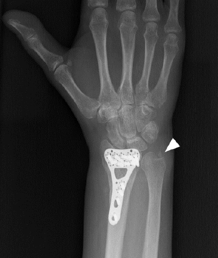
jyms-2023-01291f1.jpg
Fig. 2.
Anteroposterior radiograph shows the metal plate in the radius and the fracture of the ulnar styloid (arrowhead).
jyms-2023-01291f2.jpg
  • 1. Ricci V, Ricci C, Cocco G, Gervasoni F, Donati D, Farì G, et al. Histopathology and high-resolution ultrasound imaging for peripheral nerve (injuries). J Neurol 2022;269:3663–75.ArticlePubMedPDF
  • 2. Lee JH, Kim KJ, Baek JH. Factors affecting the occurrence of late median nerve neuropathy after open reduction and volar locking plate fixation of distal radius fracture. Orthopedics 2021;44:e367–72.ArticlePubMed
  • 3. Yeh KT, Lee RP, Yu TC, Wang JH, Liu KL, Peng CH, et al. Risk factors for carpal tunnel syndrome or trigger finger following distal radius fracture: a nationwide study. Sci Rep 2020;10:469.ArticlePubMedPMCPDF
  • 4. Ricci V, Soylu AR, Özçakar L. Artifacts and artistic facts: a visual simulation for ultrasound training. Am J Phys Med Rehabil 2019;98:521–5.ArticlePubMed
  • 5. Mezian K, Ricci V, Güvener O, Jačisko J, Novotny T, Kara M, et al. EURO-MUSCULUS/USPRM dynamic ultrasound protocols for wrist and hand. Am J Phys Med Rehabil 2022;101:e132–8.ArticlePubMed

Figure & Data

References

    Citations

    Citations to this article as recorded by  

      Figure

      JYMS : Journal of Yeungnam Medical Science