Effectiveness of orthoses for treatment in patients with spinal pain
Article information

Abstract
Spinal pain is a common patient complaint in clinical practice. Conservative treatment methods include oral medication, physical therapy, injections, and spinal orthoses. The clinical application of orthoses is debated because of potential complications associated with long-term use, such as muscle weakness and joint contracture. We reviewed the orthoses most frequently used to manage spinal pain. We review the use of soft cervical and Philadelphia collars, lumbosacral corsets, and thoracolumbosacral orthosis to manage spinal pain. Spinal orthoses can help reduce pain by protecting the muscles and joints of the injured spinal region, preventing or correcting malformations, and limiting trunk flexion, extension, lateral flexion, and rotation. The short-term use of spinal orthoses is known to improve pain and disability during the treatment period without significant adverse effects. Spinal orthoses are expected to alleviate pain and improve patients’ lifestyle.
Introduction
Spinal pain is a common patient complaint, affecting 80%–90% of individuals at least once in their lifetime [1,2]. There are various causes of spinal pain, such as spinal degeneration, trauma, inflammation, infection, and deformities. In clinical practice, spinal degeneration (herniated disc or spinal stenosis) and trauma are the most common causes of spinal pain [3-5]. To alleviate spinal pain, conservative treatments, including rest, physiotherapy (e.g., heat therapy, traction therapy, and manual therapy), injections, orthoses, and medication, are used before the surgical treatment [6-8]. Although the clinical application of orthoses is debated because of potential complications associated with long-term use, such as muscle weakness and joint contracture, its short-term use is known to improve pain and disability during the treatment period without significant adverse effects [9-11].
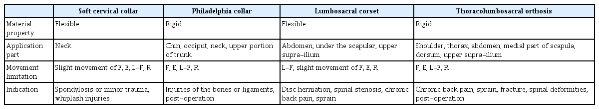
In this study, we reviewed the following types of orthoses most frequently used to manage spinal pain: soft cervical and Philadelphia collars, lumbosacral corset, and thoracolumbosacral orthosis (Table 1).
Characteristics of spinal orthoses
Soft cervical collar
The soft cervical collar is comprised of a soft foam material, a fabric covering the foam, and a Velcro strap (Fig. 1) [12,13]. The strap is mostly fastened at the back but can also be placed at the front, depending on the user’s preference. Patients wearing a soft cervical collar can experience feelings of warmth and psychological comfort owing to the fabric sheathing [14]. However, soft cervical collars cannot significantly restrict the cervical spine’s range of motion, thus falling short in providing sufficient structural support [12,14]. Therefore, they are used to manage muscle pain and spasms due to spondylosis or minor trauma and as an initial treatment for whiplash injuries. According to a previous study, the soft cervical collar is recommended to be worn for 2 weeks [11].
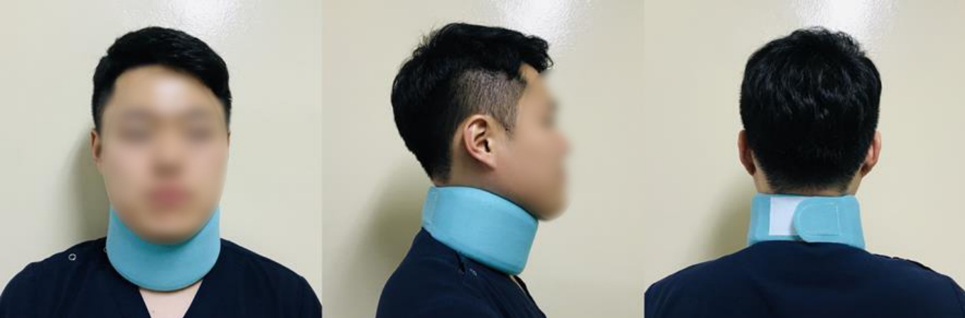
Soft cervical collar. The subject is wearing soft cervical collar.
Muzin et al. [11] reported no side effects (e.g., muscle weakness) associated with the use of soft cervical collars for fewer than 10 days. Mealy et al. [15] assessed 61 patients with acute cervical whiplash injuries following the use of the soft cervical collar for 2 weeks. The patients were divided into the following two groups: one that progressively combined exercise with use of the soft cervical collar and the other that performed exercise without use of the soft cervical collar. Eight weeks later, the visual analog scale (VAS) score from 0 to 10 (with 10 indicating unsustainable pain and 0 indicating absence of pain) and range of motion of the cervical spine (i.e., flexion, extension, lateral flexion, and rotation) were obtained. The group using the soft cervical collar reported a higher reduction in both the intensity of pain and range of motion of the cervical spine [15]. Furthermore, Rosenfeld et al. [16] investigated 97 patients with whiplash injuries by dividing them into two groups as follows: (1) those who wore soft cervical collars within 96 hours from the injury and were treated after 2 weeks and (2) those who did not use the collar and were treated using the same protocol. In addition, the VAS scores for pain were obtained for both the groups. After 6 months, the VAS scores decreased by 3 points in the group that used the soft cervical collars and only by 1.5 points in the group that did not use the soft cervical collar.
Philadelphia collar
The Philadelphia collar, which usually comprises of a solid plastic sheet, limits a greater range of movements compared to the soft cervical collar (Fig. 2) [12]. It is vertically reinforced from the chin to the manubrium in the front and shaped to cover the area from the external protuberance of the occipital bone to the upper part of the spine of the scapula at the back. The anterior part of the Philadelphia collar has a hole for tracheostomy; therefore, the user’s chin has to be aligned with its center [17,18]. Furthermore, the inner side of the Philadelphia collar is lined with a replaceable padding, which permits good hygiene and causes less irritation to the skin. The Philadelphia collar slightly reduces the load on the spine by promoting the correct posture at the cervical spine and plays a role in limiting the cervical flexion/extension, lateral flexions and rotation [18,19]. Nonetheless, some pressure may be applied on the clavicle by the Philadelphia collar. Considering that excessive pressure can cause discomfort or pressure sores, special attention is required for users with sensitive skin [18,20]. The Philadelphia collar can be used to treat injuries of the bones and ligaments in the mid-cervical spine region and for postsurgical stabilization. In addition, it can be used instead of the halo orthosis to stabilize upper cervical fractures (Jefferson and hangman’s fractures) and fractures of the odontoid process [11].
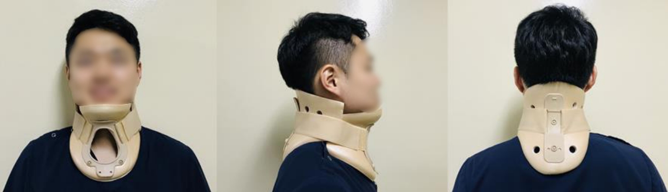
Philadelphia collar. The subject is wearing Philadelphia collar.
According to a study by Beavis [14], hard cervical collars are effective in limiting the motion of the cervical spine (level of movement reduction: flexion 69%, extension 34%, left lateral flexion 22%, right lateral flexion 34%, leftward rotation 50%, and rightward rotation 48%). Similarly, Muzin et al. [11] suggested that hard cervical collars were effective for the initial management of trauma (i.e., to prevent cervical instability). Finally, Motiei-Langroudi and Sadeghian [21] studied 11 patients with a C2 fracture who used the Philadelphia collar either until the bone completely recovered or until the neck pain disappeared. After a follow-up of 21 months, the patients reported no neurological symptoms or deficits, a mean VAS score of 2, and recovery of their lifestyle before the injury.
Lumbosacral corset
The lumbosacral corset is comprised of soft materials. It encloses the trunk and pelvis and has a string or hook to adjust the circumference. When necessary, a canvas, nylon mesh, or coil spring is used to increase its capability to provide support (Fig. 3). The posterior support column is made of semirigid or soft plastic materials and is molded to the shape of the patient’s body. It is inserted into a corset, which confers rigidity to the support, thus limiting hyperextension of the spine and reducing spinal lordosis [22-25]. In the groin region, straps can be attached to prevent the movement of the corset. The upper margin of the anterior surface of the corset is positioned 1.3 cm (1/2 inch) below the xiphoid process, and the lower margin is located 2.5–3 cm (1 inch) above the pubic symphysis. Furthermore, while the upper margin of the posterior surface is 2.5 cm (1 inch) below the inferior angle of the scapula, the lower margin is located at the most prominent part of the hip [18].
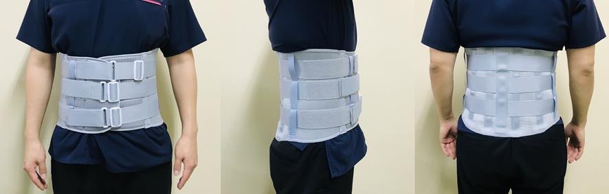
Lumbosacral corset. The subject is wearing lumbosacral corset.
The lumbosacral corset limits several movements in the frontal plane. It is less rigid at the pelvis, allowing movements in both the sagittal and transverse planes. Moreover, the corset applies pressure on the abdomen; therefore, the intraabdominal pressure increases, which reduces stress on the spine and load on the spinal disc and extensors. Furthermore, it enhances the user’s perception of proprioception. The lumbosacral corset can be used in cases of disc herniation, spinal stenosis, chronic back pain, pelvic fracture, and sprain of the lumbosacral spine [23-27].
Kim [28] investigated 69 patients who used the lumbosacral corset to treat a herniated disc or sprain in the lumbar spine region. Physical examinations (e.g., straight leg raising [SLR] and gait analysis) were performed, and the intensity of pain and clinical outcomes of using the lumbosacral corset were determined based on the criteria suggested by Stauffer and Coventry [29]. While the results of both SLR and gait analysis were “poor” in 63.77% and 59.4% of the patients, respectively, prior to use of the lumbosacral corset, improvements were noticed following its use (i.e., 84.06% and 85.50% of the patients reported a greater than “fair” result). These findings suggest that the use of the lumbosacral corset is effective in reducing pain and improving the activities of daily living. In 2001, Prateepavanich et al. [30] measured the claudication distance and pain score (VAS) in 21 patients with lumbar spinal stenosis without wearing a lumbosacral corset, and a week later with a lumbosacral corset, evaluated again and compared the results. As a result, a significant difference between the two groups, an average claudication distance was 393.2 m when wearing a lumbosacral corset and 314.6 m when not wearing a lumbosacral corset, and a mean value of pain score was 4.7 when wearing a corset and 5.9 when not wearing a lumbosacral corset. This result showed that the effects of lumbosacral corset in lumbar spinal stenosis.
Thoracolumbosacral orthosis
The thoracolumbosacral orthosis (TLSO) can be of the following two types based on the type of material used: soft and hard. In addition, it is classified based on the location and presence of structural elements as flexion adjustable, flexion/extension adjustable, flexion/extension/lateral flexion adjustable, and flexion/extension/lateral flexion/rotation adjustable. It is fabricated on the principle of three-point pressure. TLSO can be used to treat chronic back pain, sprains and fractures of the thoracic or lumbar spine, and spinal deformities, and for postsurgical management of the spine [26,31].
TLSO is widely used in current clinical practice (Fig. 4). Owing to its light and breathable mesh fabric, it provides a comfortable fit. Furthermore, ergonomically designed plastic panels provide high stability through the application of abdominal pressure and insertion of the back panels. In addition, TLSO is adjustable through the connection of two elastic straps so as to fit the shape of the patient’s body. It has a shoulder strap to prevent it from sliding out of place.
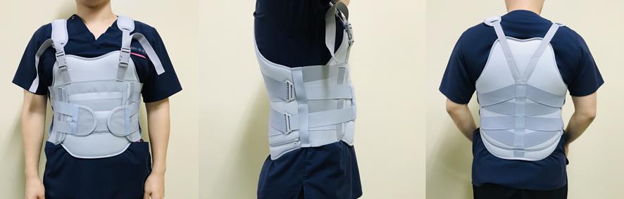
Thoracolumbosacral orthosis. The subject is wearing thoracolumbosacral orthosis.
Jacobs et al. [32] assessed 15 patients with an osteoporotic vertebral compression fracture. Following the use of a semirigid TLSO for 6 weeks, their VAS scores and quality of life (measured using the Quality of Life Questionnaire of the European Foundation for Osteoporosis) were evaluated. The outcomes were decreased mean VAS scores (i.e., from 5 to 2 points) and improvements in pain (38%), physical function (42%), social function (21%), and health perception (16%).
Conclusion
Several patients with spinal pain are encountered in clinical practice. Spinal orthoses are expected to alleviate pain and improve patients’ lifestyle. Nevertheless, studies on the clinical efficacy of orthoses are neither quantitatively nor qualitatively sufficient to reach a solid conclusion. Therefore, additional investigations are required to issue guidelines on the appropriate use of spinal orthoses. Our study is limited in that we reviewed only most commonly used orthoses, accordingly more various orthoses should be reviewed in the future study.
Notes
Conflicts of interest
No potential conflict of interest relevant to this article was reported.
Author contributions
Conceptualization: YJC, MCC; Data curation: YJC, MCC; Formal analysis: MCC; Methodology: YJC, MCC; Investigation: YJC; Resources: YJC; Supervision: MCC; Visualization: YJC, MCC; Writing-original draft: YJC, MCC; Writing-review & editing: YJC, MCC.
Additional information
The model provided written informed consent for the use and publication of his photographs.