Transient osteoporosis of the hip with a femoral neck fracture during follow-up: a case report
Article information

Abstract
We report a case of transient osteoporosis of the hip with a femoral neck fracture found during follow-up. A 53-year-old man presented with left hip pain without trauma. The pain did not improve after 2 weeks and he was brought to our hospital by ambulance. Magnetic resonance imaging (MRI) of the left hip joint showed diffuse edema in the bone marrow, which was identified by low signal intensity on T1-weighted images, high signal intensity on T2-weighted images, and increased signal intensity on short tau inversion recovery. This edema extended from the femoral head and neck to the intertrochanteric area. He was diagnosed with transient osteoporosis of the left hip. Rest gradually improved his pain; however, 3 weeks later, his left hip pain worsened without trauma. X-ray, computed tomography, and MRI results of the hip joint demonstrated a left femoral neck fracture, and osteosynthesis was performed. Differential diagnoses included avascular necrosis of the femoral head, infection, complex regional pain syndrome, rheumatoid arthritis, leukemia, and other cancers. Transient osteoporosis of the hip generally has a good prognosis with spontaneous remission within a few months to 1 year. However, a sufficient length of follow-up from condition onset to full recovery is necessary to avoid all probable complications such as fractures.
Introduction
Transient osteoporosis of the hip (TOH) is a rare disease that was first reported in 1959 in three pregnant women who had strong unilateral or bilateral hip pain [1]. TOH generally occurs in middle-aged men and pregnant women in their last trimester [2-4]. Its etiology is unknown; however, it is suspected that ischemia in the femoral head could be involved in the onset of TOH [5-7]. Differential diagnosis includes avascular necrosis of the femoral head, infection, complex regional pain syndrome, rheumatoid arthritis, leukemia, and other cancers. TOH is conventionally a disease with a good prognosis and spontaneous remission within a few months to 1 year. We report a case of TOH where a femoral neck fracture later occurred without trauma, and osteosynthesis was performed.
Case
Ethical statements: Written informed consent was obtained from the patient for this case report. This study was approved by the Institutional Review Board (IRB) of Musashino General Hospital (IRB No: 12).
A 53-year-old man presented with left hip pain without trauma that had persisted for 2 weeks. After the pain did not subside, he was brought to our hospital by ambulance. He had schizophrenia and epilepsy resulting from a previous head injury. He drank approximately 1.5 L of beer every day and had smoked 20 cigarettes per day for 37 years.
On the first admission, he had difficulty walking due to severe left hip pain. The blood examination was normal. His left hip joint demonstrated painful limitations in passive range of motion. Patrick’s test was positive.
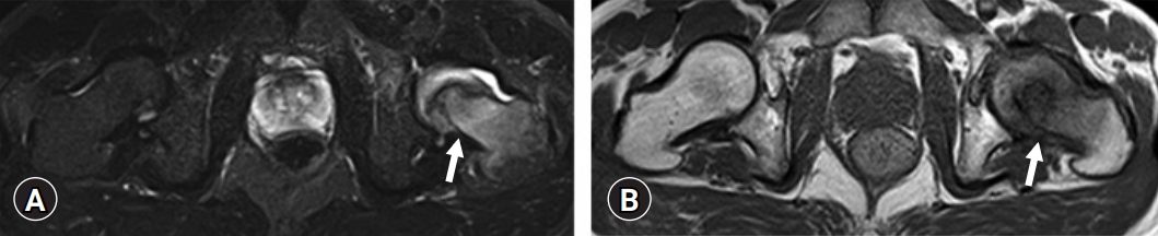
X-ray and computed tomography (CT) revealed no obvious fracture (Fig. 1). Magnetic resonance imaging (MRI) showed diffuse edema in the bone marrow, which was identified by low signal intensity on T1-weighted images, high signal intensity on T2-weighted images, and increased signal intensity on short tau inversion recovery (STIR). This edema extended from the femoral head and neck to the intertrochanteric area (Figs. 2, 3).
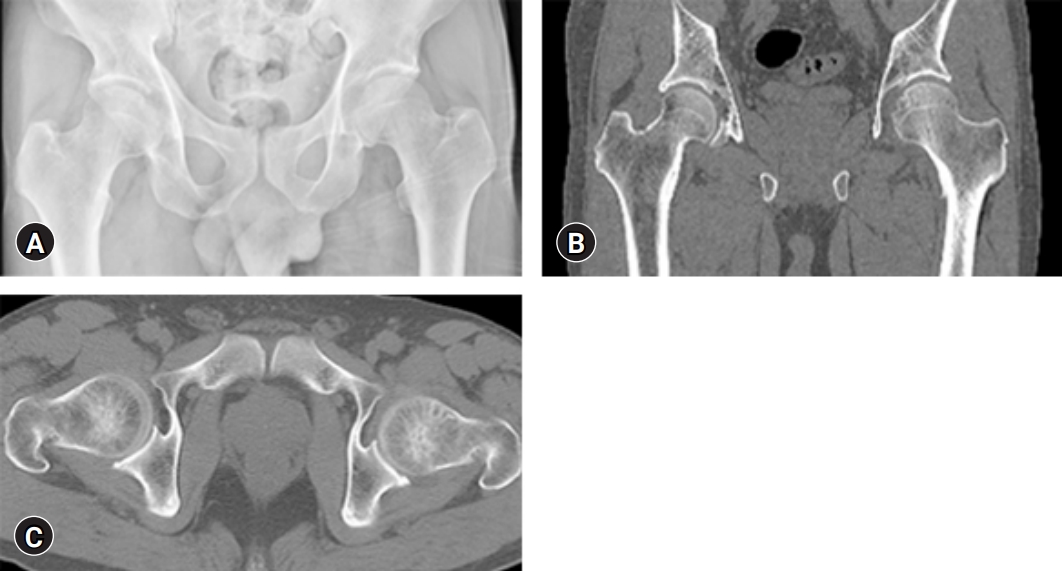
X-ray and computed tomography (CT) reveal no obvious fractures. (A) X-ray anterior view. (B) CT coronal view. (C) CT axial view.

The magnetic resonance imaging coronal views of the left transient osteoporosis of the hip. (A) Short tau inversion recovery image. (B) T2-weighted image. (C) T1-weighted image.
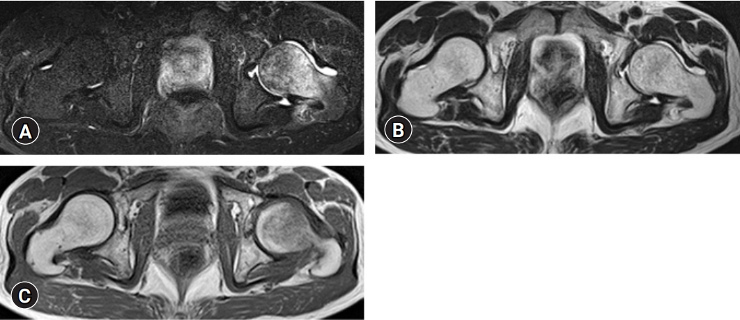
Magnetic resonance imaging (MRI) axial view of the left transient osteoporosis of the hip. MRI shows diffuse edema in the bone marrow, extending from the femoral head and neck to the intertrochanteric area. (A) Short tau inversion recovery image. (B) T2-weighted image. (C) T1-weighted image.
We prescribed rest and non-weight bearing for him; however, he did not follow our instructions due to his psychological condition. His pain gradually improved and he was discharged from our hospital using crutches.
Three weeks later, his left hip pain worsened without trauma and he visited our hospital again. X-ray, CT scan, and MRI of the hip joint demonstrated a left femoral neck fracture (Garden stage Ⅱ) (Figs. 4–6), and osteosynthesis (Prima Hip Screw System, Japan Medical Dynamic Marketing, Inc., Tokyo, Japan) was performed (Fig. 7).
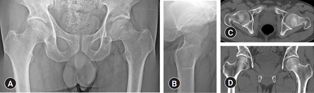
The left femoral neck fracture (arrows). It is difficult to find the fracture line of the femoral neck, by either X-ray or computed tomography (CT). (A) X-ray anterior view. (B) X-ray lateral view. (C) CT axial view. (D) CT coronal view.
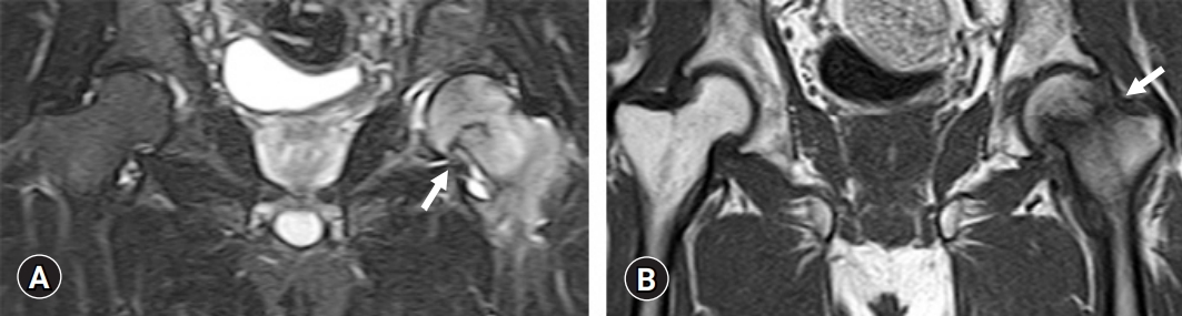
Magnetic resonance imaging coronal view of the left femoral neck fracture (arrows). (A) Short tau inversion recovery image. (B) T1-weighted image.
Magnetic resonance imaging (MRI) axial view of the left femoral neck fracture (arrows). MRI shows a clear fracture line in the left femoral neck. (A) Short tau inversion recovery image. (B) T1-weighted image.
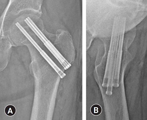
Osteosynthesis with three Prima Hip screws (Japan Medical Dynamic Marketing, Inc., Tokyo, Japan). (A) X-ray anterior view. (B) X-ray lateral view.
Discussion
TOH generally affects middle-aged men and pregnant women in their last trimester [2-4]. It is usually bilateral in pregnant women and unilateral in middle-aged men [8]. The cause and pathogenesis of TOH remain unclear; however, the most dominant theory implicates femoral head ischemia due to venous obstruction as the cause [5-7]. On examination, bone biopsies of patients with TOH showed increased numbers of erythrocytes, suggesting the presence of venous stasis [5]. Furthermore, Orth and Anagnostakos [9] reported that decreased concentrations of fibrinolytic agents and increased concentrations of thrombophilia markers in TOH patients could potentially cause venous obstruction. Risk factors for TOH include trauma, a history of steroid use, consumption of alcohol, smoking, low testosterone levels, low vitamin D levels, osteogenesis imperfecta, hypothyroidism, and hypophosphatasia [10-13]. In this case, smoking and alcohol were mentioned as risk factors.
MRI is the most powerful tool for the diagnosis of TOH and usually shows low signal intensity on T1-weighted images, high signal intensity on T2-weighted images, and increased signal intensity on STIR, reflecting bone marrow edema.
TOH typically has a good prognosis with spontaneous remission occurring within a few months to 1 year. Methods for joint preservation, including restricted weight bearing, are the first-line therapy. As alternative treatments, the use of bisphosphonates to inhibit bone resorption and the use of teriparatide as an osteoanabolic agent were reported to provide successful outcomes [14-17]. Additionally, hyperbaric oxygen therapy was reported to potentially accelerate recovery in TOH patients, although the effects were not statistically significant [18]. More recently, out of 15 TOH cases, 10 underwent hip drilling for core decompression while five underwent conservative therapy. An investigation of the time to full recovery revealed that hip drilling required a median of 5.8 weeks while conservative therapy required 48.3 weeks, demonstrating the validity of hip drilling as a treatment modality [19].
In the present case, regarding our recommendations to avoid bearing weight, the patient was nonadherent due to his poor mental health; therefore, it was assumed that the constant stress of weight bearing caused the femoral neck fracture. In fact, the fracture pattern in this case originated in the lower part of the femoral neck and is considered to be a pathological condition similar to insufficient fracture secondary to TOH. Interestingly, Hadji et al. [20] demonstrated that out of 52 TOH patients, 12.1% sustained a hip fracture. Therefore, for patients with a high likelihood of fracture, it is necessary to provide thorough education, such as instructions not to apply a load to the lower limbs during a specified period. Prophylactic osteosynthesis may also be considered for patients expected to have a high likelihood of fracture. Considering these factors, a sufficient follow-up period from condition onset to full recovery is necessary to avoid as many complications as possible.
Notes
Conflicts of interest
No potential conflict of interest relevant to this article was reported.
Funding
None.
Author contributions
Conceptualization, Supervision: YT, TM; Data curation: YT, SM, MM; Formal analysis: SM, MM, KO, YT, TM; Investigation: YT; Writing-original draft: YT; Writing-review & editing: all authors.