Isolated tubal torsion in the third trimester of pregnancy managed with simultaneous salpingectomy and cesarean section
Article information

Abstract
Isolated tubal torsion is an uncommon cause of acute abdomen in pregnancy. Tubal torsion may occur in the absence of adnexal disease. Diagnosing tubal torsion is especially difficult in pregnancy because no precise preoperative radiological and biochemical investigations have been conducted. Most patients are diagnosed during surgery. Here, I present a case of isolated tubal torsion in a pregnant woman at 35 weeks and 6 days of gestation that was managed with salpingectomy and cesarean section simultaneously.
Introduction
Isolated tubal torsion is an extremely rare event with a reported incidence of 1 in 1.5 million women. Most of the cases occur in nonpregnant women but approximately 12% of isolated tubal torsion were diagnosed during pregnancy [1]. The first case of tubal torsion in women was reported in 1890 by Bland-Sutton [2]. Diagnosing isolated tubal torsion is difficult because of the wide range of differential diagnoses including many other emergency causes for abdominal pain such as ovarian torsion, appendicitis, pelvic inflammatory disease, ectopic pregnancy, renal colic, and diverticulitis. The predisposing factors are either extrinsic abnormalities, including paratubal mass, peritubal adhesions, and uterine enlargement compressing the fallopian tubes, or intrinsic tubal abnormalities, including hydrosalpinx, tubal ligation, or endometriosis [3,4]. The risk increases in pregnancy particularly in the presence of the above-mentioned predisposing factors.
Here, I report a case of isolated tubal torsion in a pregnant woman at 35 weeks and 6 days of gestation that was managed with salpingectomy and cesarean section simultaneously.
Case
A 36-year-old, gravida 2, para 1, patient at 35 weeks and 6 days of gestation was referred to our hospital because of acute right abdominal pain that began 2 days prior to visit. However, she had no vomiting, diarrhea, constipation, or fever. In the right lower quadrant, direct tenderness without rebound tenderness was the only abnormal finding in her physical examination. She had no medical history and the surgical history did not reveal any abnormalities except that she gave birth to a full-term, healthy baby boy by cesarean section 2 years earlier. Her vital signs at the time of admission were normal and the laboratory tests showd the following results: hemoglobin 10.8 g/dL (range, 12-15.9 g/dL), white blood cells 9,330/µL (range, 4,000-10,000/µL), and platelets 213,000/µL (range, 140,000-400,000/µL), with C-reactive protein level increased to 42 mg/L (range, 0-5 mg/L). The non-contrast-enhanced abdominopelvic computed tomography (CT) performed in the secondary hospital prior to referral to our hospital revealed a homogeneous cystic mass measuring 44×25 mm in the right lower quadrant; no other abnormal findings were observed (Fig. 1A).
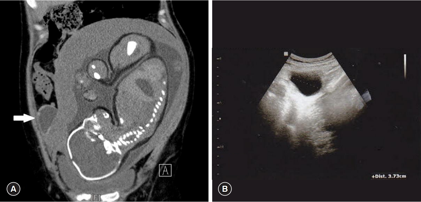
(A) Abdominopelvic computed tomography shows a 44×25 mm sized homogenous cystic mass occupies the right lower quadrant. (B) Transabdominal ultrasonography shows a 37 mm sized hypoechoic cystic mass in the right lower quadrant.
Ultrasonography performed in our hospital revealed a single live fetus at 36 weeks of gestation with normal amniotic fluid and placenta as well as a 37-mm sized hypoechoic cystic mass in the right lower quadrant (Fig. 1B). The nonstress test did not provoke any uterine contractions and showed normal accelerations and variability of the fetal heart rate. Because the pain in her right lower abdomen did not subside and tended toward exacerbation within 24 hours of admission, I suspected right adnexal torsion, appendicitis, or rupture of right ovarian cyst. Exploratory laparotomy was performed to prevent complications such as tissue necrosis caused by adnexal torsion or panperitonitis caused by appendicitis. Given the 36 weeks of gestation and normal fetal status as well as the history of cesarean section I decided to perform a simultaneous cesarean section. A healthy baby girl weighing 2,500 g was born. There was no abnormal finding such as adhesion during cesarean section but torsion of midportion of the right fallopian tube and necrosis measuring 6×4 cm size was observed (Fig. 2A). The cystic mass observed on CT scan and ultrasonography was hydrosalpinx which was thought to be the result of ischemic or traumatic tubal injury due to torsion. The contralateral fallopian tube both ovaries and the appearance and size of the appendix were normal. A right salpingectomy was performed and the operative specimen revealed an ovoid shaped, purple-brown colored, diffusely congested fallopian tube (Fig. 2B). The surgery was completed without any complication. The postoperative recovery of the patient and follow-up of her baby were uneventful. A histological examination revealed that the wall of the fallopian tube was severely edematous and congested.
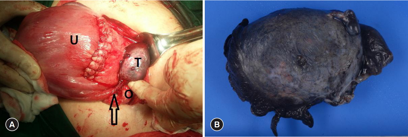
(A) Intraoperative findings: a right ischemic fallopian tubal mass (T) and a grossly normal ovary (O) are beside uterus (U). The torsion of the right fallopian tube (arrow) was observed at midportion. (B) Operative specimen: a 6×4 cm sized, ovoid shaped, purple-brown colored, diffusely congested fallopian tube.
Discussion
Isolated tubal torsion is one of various conditions accounting for lower abdominal pain that usually affects reproductive women. The condition is extremely rare (i.e., 1 in 1.5 million women), and has been described as a rare cause of acute abdomen in pregnancy. As far as I know, approximately 30 cases of isolated tubal torsion during pregnancy seem to have been reported in English literatures [5-11]. The lack of specific clinical presentation, laboratory findings, and imaging features makes it difficult to identify an isolated tubal torsion preoperatively and subsequently delays prompt surgical intervention to prevent irreversible vascular changes in the fallopian tube [12].
Torsion generally occurs in abnormal fallopian tubes; however, it might also develop in normal ones. Structural abnormalities such as hematosalpinx, hydrosalpinx, cyst of Morgagni, previous tubal ligation or surgery, peritubal adhesions, and tubal or ovarian neoplasm are believed to play a role in the development of torsion. Moreover, hormonal medications which result in hypermobility or tubal spasm by affecting the normal physiology, trauma to the fallopian tube and varicose veins in the mesosalpinx are also reported to be other etiological factors [13].
In this case, the only obvious etiological factor for the development of the twisted fallopian tube was assumed to be pregnancy which can play a role as a rotational force resulting from changes in the abdominal cavity and uterine size. Any adhesions related to the previous cesarean section could also be a possible etiological factor, but there was no abnormal finding in this case. A paratubal cyst could not be diagnosed on pathological examination. Tubal torsion occurs more commonly on the right side than on the left. Presumably, the possibility of developing torsion on the right side is higher because of the movement of the appendix and small bowel on the right side while the mesentery of the sigmoid colon is attached to the left side [14].
Imaging findings in isolated tubal torsion are nonspecific and clinical correlation is necessary. In the presence of acute pain, the ultrasonographic findings of a dilated tube with a normal-appearing ipsilateral ovary should point to the possibility of isolated tubal torsion. Ultrasonographic signs of a dilated fallopian tube are a hyperechoic wall a folded configuration and foci of echogenicity protruding into the lumen, and a tapering end broad toward the uterine cornua. In such situations, the fallopian tube is examined for a whirlpool mass in close proximity. This mass is usually smaller and less obvious than those seen in ovarian torsion [15]. In this case, the patient underwent CT scan to evaluate acute pain at another hospital. However, performing a CT scan on the mother is not appropriate because it exposes the fetus to high radiation. Magnetic resonance imaging is considered to be more appropriate than CT scan if additional imaging test is required after first performing an ultrasound.
Surgery by laparoscopy or laparotomy is the standard treatment for isolated tubal torsion and leads to a definitive diagnosis. Laparoscopy remains the standard procedure for diagnosis and treatment as early diagnosis may allow detorsion/salvage of the tube in nonpregnant patients and patients in early pregnancy. In view of technical difficulties of the laparoscopic access to adnexa, laparotomy is generally preferred in advanced gestation [5]. In addition, most surgeons consider laparotomy appropriate in advanced third-trimester pregnancy in view of the possible option of delivering the fetus as seen in this patient. A patient with tubal torsion without any ischemic damage can be successfully treated with tubal detorsion which is preferred in the reproductive age group. If the tube is gangrenous, a malignancy can be suspected, and if the tube is diseased or if the patient does not pregnancy in the future, salpingectomy is preferred. In this case, since the tube was necrotic and no further pregnancies were planned, salpingectomy was performed.
In conclusion, isolated tubal torsion in pregnancy is an extremely rare cause of acute abdomen. However, it should be included in the differential diagnosis of acute abdomen in pregnancy. Early decision regarding surgical intervention will prevent obstetric complications and may allow preservation of the tube.
Acknowledgements
This work was supported by research grant of Wonkwang University in 2018.
Notes
No potential conflicts of interest relevant to this article was reported.