E-Submission
Indexed in: ESCI, Scopus, PubMed,
PubMed Central, CAS, DOAJ, KCI
PubMed Central, CAS, DOAJ, KCI
FREE article processing charge

Articles
- Page Path
- HOME > J Yeungnam Med Sci > Volume 35(1); 2018 > Article
-
Case Report
Multi-vessel intractable coronary spasm development in a patient with aborted sudden cardiac death: a case study with intravascular ultrasound findings -
Sungsoo Cho
 , Tae Soo Kang
 -
Yeungnam University Journal of Medicine 2018;35(1):121-126.
DOI: https://doi.org/10.12701/yujm.2018.35.1.121
Published online: June 30, 2018
Division of Cardiovascular Medicine, Department of Internal Medicine, Dankook University Hospital, Dankook University College of Medicine, Cheonan, Korea
- Corresponding Author: Tae Soo Kang, Division of Cardiovascular Medicine, Department of Internal Medicine, Dankook University Hospital, Dankook University College of Medicine, 201, Manghyang-ro, Dongnam-gu, Cheonan 31116, Korea Tel: +82-41-550-7690, Fax: +82-41-556-0524 E-mail: neosoo70@dankook.ac.kr
• Received: January 17, 2018 • Revised: January 29, 2018 • Accepted: February 9, 2018
Copyright © 2018 Yeungnam University College of Medicine
This is an Open Access article distributed under the terms of the Creative Commons Attribution Non-Commercial License (http://creativecommons.org/licenses/by-nc/4.0/) which permits unrestricted non-commercial use, distribution, and reproduction in any medium, provided the original work is properly cited.
- 4,368 Views
- 53 Download
- 1 Crossref
Abstract
- Coronary spasm generally occurs in patients with minimal atherosclerotic plaque lesion, and it has a rather favorable prognosis. However, in some cases, coronary spasm may induce myocardial infarction and even sudden cardiac death (SCD). Here, we report a case in which multi-vessel intractable coronary vasospasm suddenly occurred in a diffuse atherosclerotic lesion after percutaneous coronary intervention (PCI) in a patient with aborted SCD. We identified the characteristics of the spasm portion in intravascular ultrasound (IVUS) images and conducted percutaneous cardiopulmonary bypass support-PCI with stenting as treatment. Intima and media thickening and a large attenuated plaque burden with rupture were identified in IVUS images at the obstructive spasm portion.
- Coronary spasm generally occurs at a normal or minimally narrowed lesion on coronary angiography in relatively young patients, and some atherosclerotic plaques usually present at focal spasm sites on intravascular ultrasound (IVUS) [1,2]. Most coronary spasms have a favorable clinical outcome because they respond well to medical therapy [3]. However, in some cases, coronary vasospasm may induce an acute myocardial infarction (AMI) and even sudden cardiac death (SCD) [4]. We describe a case in which multi-vessel intractable coronary spasm suddenly occurred in a diffuse severe coronary lesion after percutaneous coronary intervention (PCI) in a patient with aborted SCD. The site of the spasm was not related to the PCI-treated portion. We identified the characteristics of the refractory coronary spasm portion in IVUS images and treated the patient with percutaneous cardiopulmonary bypass support (PCPS)-PCI with stenting.
INTRODUCTION
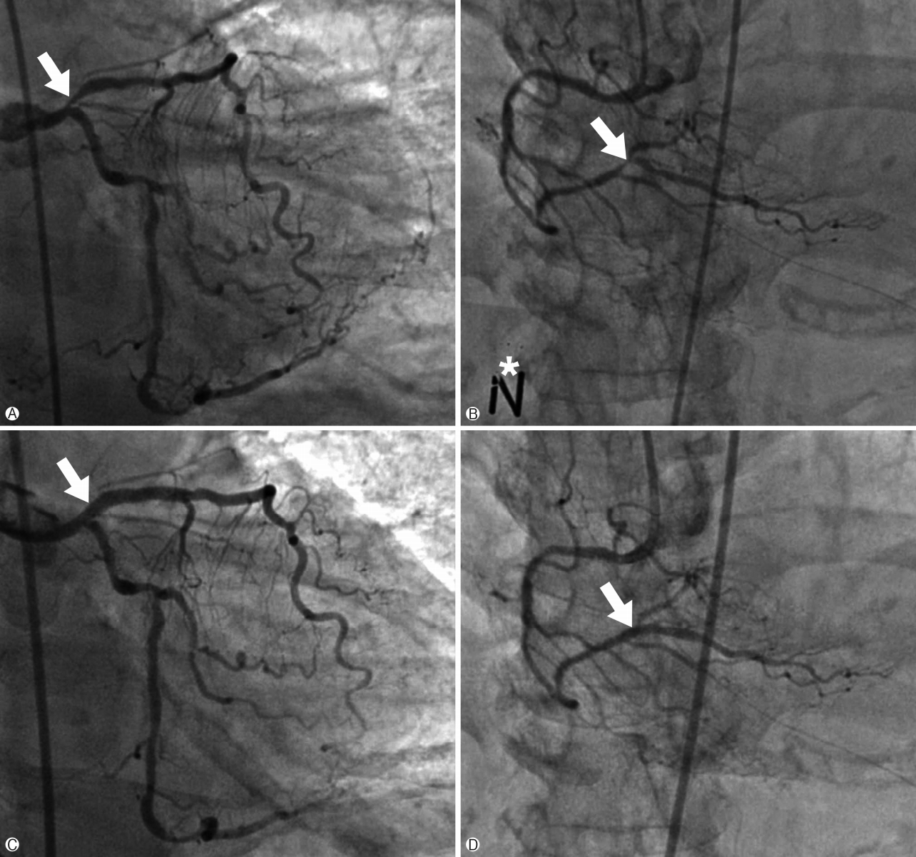
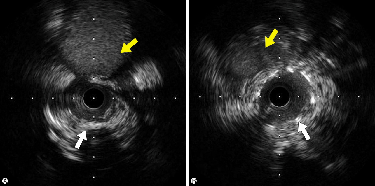
- A 72-year-old female patient was referred from a local hospital because of a sudden cardiac arrest. Cardiopulmonary resuscitation (CPR) was performed for 7 min, and the patient was resuscitated. After being transferred, she was monitored at the intensive care unit and evaluated to determine the cause of the sudden cardiac arrest. Coronary angiography revealed a significant stenosis extending from the distal left main to the proximal left anterior descending (LAD) coronary artery (Fig. 1A). The right coronary artery (RCA) showed a significant diffuse stenosis. The most severe discrete stenosis was present at the distal RCA to the posterior lateral (PL) branch lesion (Fig. 1B). We performed PCI with stenting at the short segment of the distal RCA to the PL branch (Fig. 1D). Then, we conducted an IVUS-guided PCI with stenting from the distal left main to the proximal LAD crossing over to the left circumflex (LCx) coronary artery (Fig. 1C). The IVUS image showed an eccentric echodense plaque with severe luminal narrowing at the proximal LAD, and no severe stenosis or plaque rupture was seen at the proximal LCx coronary artery (Fig. 2). After the procedure, the patient’s blood pressure gradually decreased, and she suddenly developed an atrioventricular block. We inserted a temporary pacemaker and started vasopressor therapy. Another coronary angiography showed total occlusion of the proximal RCA, causing severe spasm (Fig. 3A). We injected nicorandil, which partially resolved the spasm, and the patient’s blood pressure was elevated, and her heart rhythm was converted to sinus rhythm. IVUS of the RCA revealed diffuse intima and media thickening with a large amount of atheromatous plaque at the mid-RCA level. In the spasm portion of the proximal RCA, there was a large area of attenuated plaque and prominent intima thickening with rupture (Fig. 3B). Therefore, we performed PCI with stenting at the proximal RCA plaque rupture lesion (Fig. 3C). After stenting, the RCA flow was well maintained, and the patient was stabilized. We observed the patient for several minutes; however, her blood pressure suddenly decreased, and she again developed a pacing rhythm. We performed an RCA coronary angiography again, and the deployed stent remained patent and the flow was maintained well. We performed a left coronary angiography, which revealed total occlusion of the LCx coronary artery (Fig. 4A). We injected nicorandil and the flow was fully resolved abruptly; however, the patient again developed a spasm(Fig. 4B). Her blood pressure was continuously low, and she developed ventricular fibrillation. We performed CPR and applied PCPS, and the patient was stabilized thereafter. We conducted left coronary angiography to confirm the patency of the LCx coronary artery. However, coronary spasm again developed at the mid-RCA (Fig. 5A) and it could not be resolved despite multiple injections of nicorandil (Fig. 5B). We deployed a stent at the mid-RCA to mechanically stop the spasm. The flow was recovered after stenting (Fig. 5C). After 7 days, the patient could be weaned from the PCPS and she was discharged 1 month later. Three months later, she was admitted for a transient ischemic attack. During the hospital stay, she experienced a gastrointestinal bleeding event and heart failure aggravation. We stopped the diltiazem medication, which was the cause of reduced heart function. On the next day, she had a sudden cardiac arrest again and was resuscitated after a 5-min CPR. We intravenously injected nitrate and performed coronary angiography. All stents were patent, and no other lesion progression was found. The patient recovered and was discharged with prescriptions of clopidogrel, ramipril, statin, diltiazem, nicorandil, and isosorbide dinitrate.
CASE
- This case is clinically significant because it demonstrates that multi-vessel intractable coronary spasm can occur at a diffuse atherosclerotic lesion during the evaluation of SCD without a provocation test and confirms the IVUS imaging findings of the spasm-involved vessel.
- Coronary spasm as a cause of SCD is rare; however, there is also a certain group of patients with a very active resistant form of coronary spasm. As in our case, some reports have demonstrated that a spasm can be mechanically broken through stent implantation, when the coronary spasm was resistant to drugs and has caused serious hemodynamic collapse [5].
- There were several studies of intracoronary imaging in patients with coronary spasm. The characteristics of IVUS findings in the coronary spasm portion were diffuse intima thickening with a fibrous-dominant coronary plaque burden [6]. Ota et al. reported the IVUS findings from a case of ST-segment elevation myocardial infarction caused by coronary spasm. The IVUS findings at the spasm site showed diffuse intima and media thickening immediately after thrombectomy, which disappeared after nitrate administration [7]. Park et al. reported on a young patient who experienced two episodes of sudden cardiac arrest due to intractable coronary spasm and was successfully treated with stenting after IVUS evaluation. The IVUS of this case revealed a huge amount of atherosclerotic plaque burden with a small luminal area, minimal plaque rupture, and an intraluminal thrombus [8]. In our case, consecutive multi-vessel intractable coronary spasm developed in an old patient who survived SCD. The IVUS findings of the RCA after the spasm event revealed diffuse atheromatous plaque with intima and media thickening at the mid-RCA, and a large attenuated body of plaque and prominent intima thickening with rupture at the proximal RCA. Our case exhibited several of the previously reported IVUS findings of coronary artery spasm, including intima and media thickening, a large attenuated plaque burden, and minimal plaque rupture. Recently, an optical coherence tomography study reported that intima tear, erosion, and thin-cap fibroatheroma were identified in most segments causing coronary artery spasm in patients with acute coronary syndrome, but not in those with chronic stable variant angina [9]. Hence, patients with coronary spasm causing AMI or SCD have a more vulnerable plaque component than patients with coronary spasm causing stable or unstable angina. The degree of spasm could be related to the amount of plaque burden and plaque instability. A more aggressive and larger-scale intravascular imaging study will be needed in groups with coronary spasm causing AMI or SCD.
- In conclusion, we report on a patient with multi-vessel intractable coronary spasm who experienced SCD and was successfully treated with PCPS-PCI with stenting. The IVUS of the spasm portion revealed diffuse intima and media thickening, a large attenuated plaque burden, and prominent intima thickening with plaque rupture.
DISCUSSION
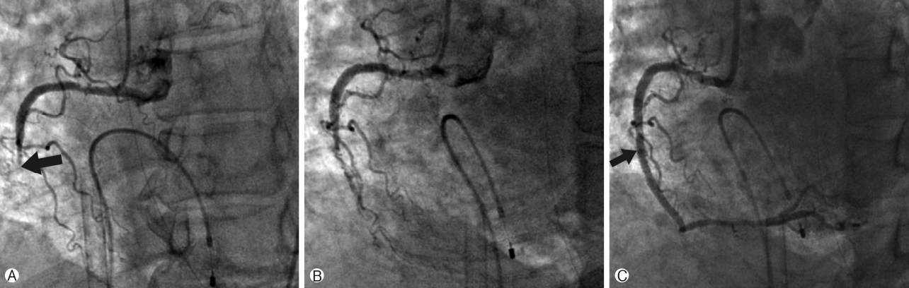
Fig. 1.Coronary angiography. (A) Initial left coronary angiography showing the significant stenosis of proximal LAD artery ostial lesion (white arrow). (B) Initial right coronary angiography showing the discrete significant stenosis of distal RCA (white arrow). *Coronary angiography after intracoronary nitrate injection. (C) Left coronary angiography after PCI and stenting at proximal LAD (white arrow). (D) Right coronary angiography after PCI and stenting at distal RCA (white arrow). LAD, left anterior descending; RCA, right coronary artery; PCI, percutaneous coronary intervention.

Fig. 2.Intravascular ultrasound. (A) Pre-intervention IVUS showing the eccentric echodense plaque with severe luminal narrowing at proximal LAD (white arrow), and there was not seen severe stenosis or plaque rupture at proximal LCx coronary artery (yellow arrow). (B) Post-intervention IVUS from distal LM to proximal LAD (white arrow: LAD and yellow arrow: LCx). LAD, left anterior descending; LCx, left circumflex; IVUS, intravascular ultrasound; PCI, percutaneous coronary intervention.

Fig. 3.Right coronary angiography and IVUS after spasm development. (A) Total occlusion of proximal RCA due to severe spasm (white arrow). (B) IVUS after resolved spasm after intracoronary nicorandil injection shows diffuse intima- and media-thickening with large amount of atheromatous plaque (yellow arrow) at mid RCA, and large attenuated plaque and prominent intima-thickening with rupture (white arrow) at proximal RCA. (C) PCI and stenting at proximal RCA (white arrow). RCA, right coronary artery; IVUS, intravascular ultrasound; PCI, percutaneous coronary intervention.
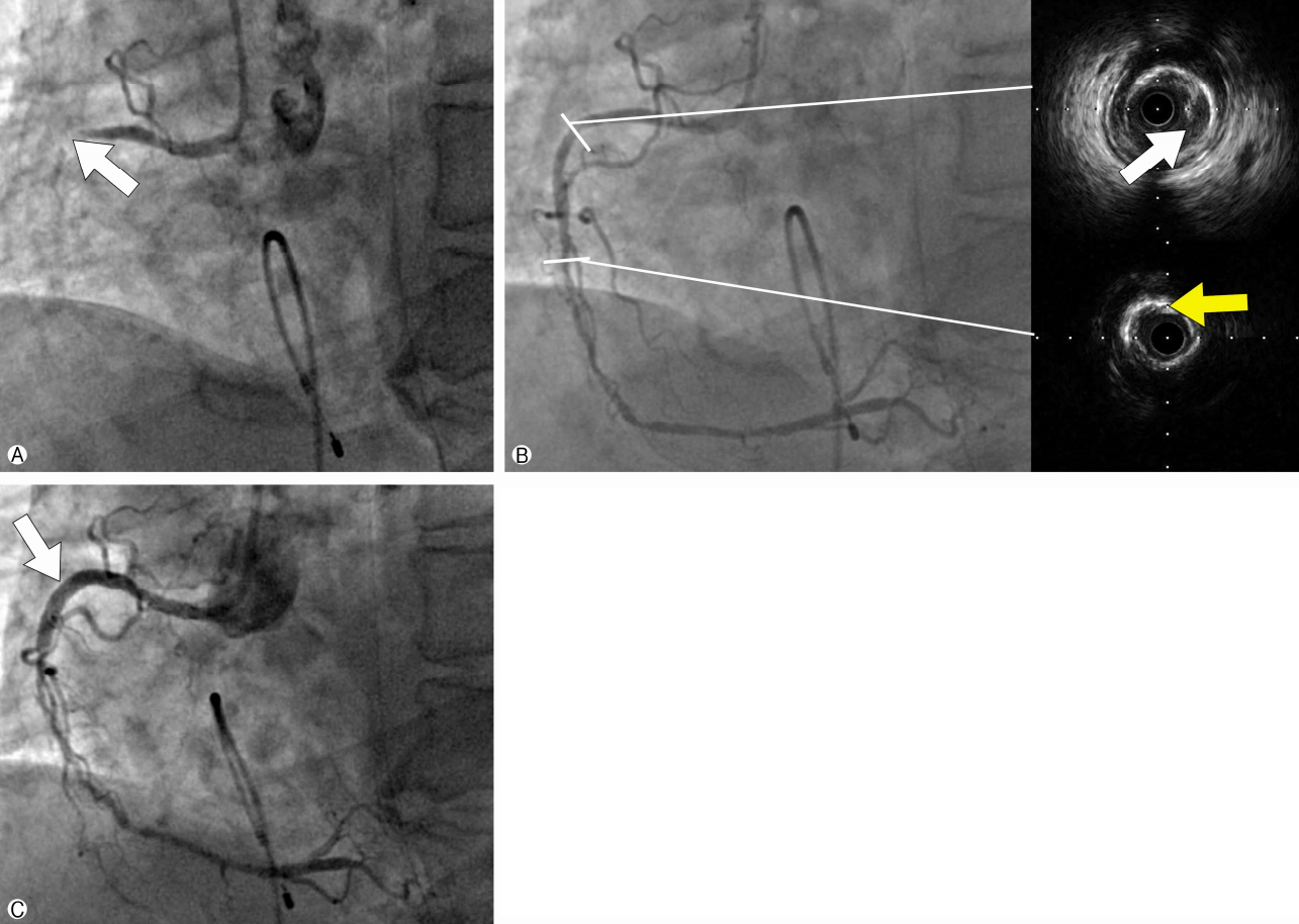
Fig. 4.Left coronary angiography after shock and recurrence AV block. (A) Total occlusion of proximal LCx artery due to severe spasm(white arrow). (B) Spasm resolved after intracoronary nicorandil injection (white arrow). AV, atrioventricular; LCx, left circumflex.
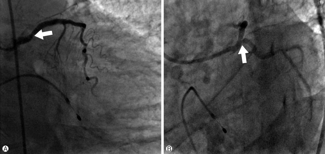
Fig. 5.Right coronary angiography after PCPS. (A) Total occlusion of mid RCA due to severe spasm (black arrow). (B) Spasm not fully resolved after several intracoronary nicorandil injections. (C) Mechanically breaking the spasm by stenting at mid RCA (black arrow). PCPS, percutaneous cardiopulmonary bypass support; RCA, right coronary artery.
- 1. JCS Joint Working Group. Guidelines for diagnosis and treatment of patients with vasospastic angina (Coronary Spastic Angina) (JCS 2013). Circ J 2014;78:2779–801.ArticlePubMed
- 2. Yamagishi M, Miyatake K, Tamai J, Nakatani S, Koyama J, Nissen SE. Intravascular ultrasound detection of atherosclerosis at the site of focal vasospasm in angiographically normal or minimally narrowed coronary segments. J Am Coll Cardiol 1994;23:352–7.ArticlePubMed
- 3. Yasue H, Takizawa A, Nagao M, Nishida S, Horie M, Kubota J, et al. Long-term prognosis for patients with variant angina and influential factors. Circulation 1988;78:1–9.ArticlePubMed
- 4. Myerburg RJ, Kessler KM, Mallon SM, Cox MM, deMarchena E, Interian A Jr, et al. Life-threatening ventricular arrhythmias in patients with silent myocardial ischemia due to coronaryartery spasm. N Engl J Med 1992;326:1451–5.ArticlePubMed
- 5. Bhagwat A, Mukhedkar S. Severe generalized resistant spasm of the right coronary artery causing hemodynamic collapse after stenting. JACC Cardiovasc Interv 2015;8:e199–200.ArticlePubMed
- 6. Miyao Y, Kugiyama K, Kawano H, Motoyama T, Ogawa H, Yoshimura M, et al. Diffuse intimal thickening of coronary arteries in patients with coronary spastic angina. J Am Coll Cardiol 2000;36:432–7.ArticlePubMedPMC
- 7. Ota H, Kawase Y, Kondo H, Miyake T, Kamikawa S, Okubo M, et al. A case report of acute myocardial infarction induced by coronary spasm. Intravascular findings. Int Heart J 2013;54:237–9.ArticlePubMed
- 8. Park YM, Kang WC, Shin KC, Han SH, Ahn T, Choi IS, et al. Repeated sudden cardiac death in coronary spasm: is IVUS helpful to decide treatment strategy? Int J Cardiol 2012;154:e57–9.ArticlePubMed
- 9. Shin ES, Ann SH, Singh GB, Lim KH, Yoon HJ, Hur SH, et al. OCT-defined morphological characteristics of coronary artery spasm sites in vasospastic angina. JACC Cardiovasc Imaging 2015;8:1059–67.ArticlePubMed
References
Figure & Data
References
Citations
Citations to this article as recorded by 
- Intractable right coronary artery spasm in the early postoperative period after heart transplantation: a case report
Jaewook Chung, Jeehoon Kang, Hae-Young Lee, Suk Ho Sohn, Ho Young Hwang, Hyun-Jai Cho
Korean Journal of Transplantation.2022; 36(2): 154. CrossRef
 PubReader
 ePub Link
 Cite
Cite